


INTRODUCTION
Although menopause involves a natural change in the female body, many studies have reported that loss of estrogen due to menopause can cause negative outcomes, such as obesity, metabolic disease, inflammation, osteoporosis, or skeletal muscle atrophy1, 2, 3. Because the female body consists of more fat and less muscle mass than the male body, skeletal muscle mass retention is the most important aspect for metabolic homeostasis, independent activity and quality of life in menopausal and postmenopausal females3, 4.
Hormone replacement therapy (HRT) has been closely associated with an improvement in metabolic health, and the maintenance of skeletal muscle mass and strength in menopausal, and postmenopausal women3. However, the various health risks of long-term HRT use have been seriously contested, and many researchers have begun to investigate the effect of alternative HRT, such as regular exercise and natural resources on inhibition of menopause-related risk factors. Numerous studies have demonstrated that various types of exercise (e.g. swimming, walking, resistance training, and Pilates) can prevent or improve deleterious effects, including skeletal muscle atrophy, on menopausal women4, 5. However, the molecular mechanisms underlying this have not yet been defined.
Classically, resistance exercise is more effective than aerobic exercise for maintenance of skeletal muscle mass through increasing protein synthesis to a greater extent than degradation, in both young and older populations6, 7, 8. According to these studies, aerobic exercise has minimal effect on skeletal muscle mass and is mainly associated with improvements in aerobic capacity, such as mitochondrial biogenesis, oxidative enzyme activity, and metabolic homeostasis. However, recent studies have suggested that aerobic exercise can also induce skeletal muscle hypertrophy through induction of skeletal muscle protein synthesis, and reduction of proteolytic systems9, 10.
An important mechanism controlling the synthesis of muscle proteins is the mammalian target of rapamycin (mTOR)11, 12, 13, 14. Resistance exercise activates mTOR and this mainly results in an increase in type II fiber (also known as fast type fiber) protein synthesis7, 13. In contrast, endurance exercise has no effect on muscle protein synthesis through the alteration of mTOR activity13,15. mTOR has a well-known positive downstream target, namely p70 ribosomal protein S6 kinase (p70S6K). It is the most well-defined effector of skeletal muscle hypertrophy through the regulation of cell growth and protein synthesis7, 11. On the other hand, tuberin (TSC) has been identified as a negative regulator of mTOR, which promotes inhibition of p70S6K activity16. In addition, activation of TSC may inhibit mTOR signaling and suppress resistance exercise-induced muscle protein synthesis7. Likewise, this signaling pathway has been considered to have a most important role in the regulation of skeletal muscle protein synthesis.
Another important mechanism for the regulation of muscle protein is the ubiquitin- proteasome system. Atrogin-1/MAFbx (atrophy gene-1/muscle atrophy F-box) and MuRF1 (muscle ring-finger protein 1) are muscle-specific ubiquitin E3-ligases and known to be required for muscle protein degradation6, 17. Therefore, many studies have tried to define a correlation between exercise and their expression level, but the effect of aerobic exercise on the ubiquitin proteasome system have shown conflicting results.
The purpose of this study was to investigate whether aerobic exercise alters skeletal muscle protein-related TSC2, p70S6K or E3-ligases in high-fat-induced ovariectomized (OVX) rats, as a model of menopause (increased body fat and estrogen deficiency), in the type II-rich plantaris muscle. Therefore, we used a long-term high-fat diet (HFD) with ovariectomy, to mimic the serious metabolic problems that are common in post-menopausal women. We hypothesized that the level of TSC2 would be down-regulated in high-fat-induced OVX rat plantaris muscles with 8 weeks of aerobic exercise, and that this mTOR-positive phenomenon would be associated with the activation of p70S6K and the reduction of ubiquitin ligases expression.
METHODS
Human and animal rights and informed consent
All animal experiments were approved by the Institutional Animal Care and Use Committee (IACUC) of Ewha Womans University, Seoul, Korea. Permit Number: 14-038.
Animal care and treatment.
Female Sprague-Dawley (SD) rats (206.19 ┬▒ 6.2 g), aged 8 weeks, were obtained from Central Lab Animal (Seoul, Korea). Animals were housed in an air-conditioned room at 23 ┬▒ 1Ōäā, 64.1% relative humidity, with a 12-hour light/dark cycle. The ovaries of all rats were removed surgically to induce a postmenopausal status and a 1-week recovery period was given. After the recovery period, the rats were randomly divided into two treatment groups: (1) high-fat diet-sedentary (HFD) and (2) high-fat diet-exercise (HFD+EX). The high-fat diets were prepared daily, for 8 weeks, using premade diets. The experimental diets were purchased from Research Diet Inc. (New Brunswick, NJ, USA). The high-fat diet contained 45% of energy as fat, derived from corn oil and lard (225:1598) (Product# D12451).
Exercise protocol
After a 1-week recovery period, the animals were acclimatized to running on a treadmill for 15 minutes, 8 m/min, at 0┬░ inclination for one day. After this, the animals were regularly trained 5 times per week, for 8 weeks, and the training started at 1000 hours. From week 1 to week 4, the animals ran on a treadmill for 30 minutes, at 15 m/min with 0┬░ inclination. Subsequently, from week 5 to week 8, the training progressed to 40 minutes and 18 m/min with 0┬░ inclination. The exercise training protocol used a modified version taken from previous studies18, 19. All training rats were restrained from training 24 hours before sacrifice.
Sacrifice and Dissection
The rats were suffocated with CO2 and the plantaris muscles were freshly dissected, trimmed, dried with filter paper, weighed and the weights recorded using an electronic balance scale (OHAUS, NJ, USA). The muscles were snap-frozen in liquid nitrogen or preserved in 4% phosphate buffered formalin for further studies.
Western blot
Tissues were lysed and homogenized in ice-cold RIPA buffer (50 mM Tris pH 7.5, 150 mM NaCl, 1% Triton X-100, 0.1% SDS, 0.5% Sodium deoxycholate, 2 mM EDTA, complete protease inhibitor, and phosphatase inhibitor cocktails). The tissue extracts were then centrifuged at 13,000 rpm for 15 min at 4Ōäā. Subsequently, the protein in the supernatant was quantified using a Bradford protein assay kit (Bio-Rad, Hercules, CA) and 80 ╬╝g of total protein were resolved on 6%-10% SDS-PAGE gel (90 V, 25Ōäā, 20 min, after 150 V, 25Ōäā, 1 hour) and transferred to a nitrocellulose membrane (25 V, 25Ōäā, 7 min). All the blots were incubated with Ponceau S (Sigma, St. Louis, MO) to ensure equal loading in all lanes (data not shown). To detect target proteins, the membranes were blocked with 5% skim milk in Tris-buffered saline (TBS) with 0.1% Tween 20 (0.1% TBST) for 1 hour, at room temperature, and subsequently incubated with total-AMPK (#2532), phospho-AMPK╬▒ (Thr172) (#2535), total-p70S6K (#9202) (polyclonal rabbit antibody, 1:1000), phospho-p70S6K (Thr389) (#9206) (monoclonal mouse antibody, 1:1000) (Cell Signaling, Beverley, MA), MAFbx/Atrogin-1 (sc- 33782) (polyclonal rabbit antibody, 1:2000) (Santa Cruz Biotechnology, Santa Cruz, CA), MuRF1 (ab77577) (polyclonal rabbit antibody, 1:1000) (Abcam, Cambridge, UK), and actin (A2066) (polyclonal rabbit antibody, 1:5000) (Sigma, St. Louis, MO, USA) antibodies overnight at 4Ōäā, in 3%-5% BSA in 0.1% TBST. Subsequently, the membranes were washed three times for 5 min each in 0.1% TBST, after which they were incubated for 1 hour with anti-mouse or rabbit IgG horse-radish peroxidase-linked secondary antibody (1:2500) (Cell signaling). The membranes were then washed as described above, after which West Femto Stable Peroxide Buffer (Thermo Fisher Scientific, Waltham, MA, USA) was applied, according to the manufacturerŌĆÖs instructions, to develop a signal that was subsequently detected using the Chemiluminescence imaging system (ATTO, Tokyo, Japan) and quantified using densitometry Image J software (NIH, USA). The target protein levels were then normalized against the actin protein levels.
Immunohistochemical staining
The plantaris muscles were directly placed in a formalin solution (Sigma, St. Louis, MO). Cross-sections were cut from the mid-belly region of each muscle. Formalin-fixed paraffin-embedded sections (5 ╬╝m) were deparaffinized, hydrated and antigen retrieval was performed using xylene. The tissue was permeabilized with 0.02% Triton X-100 in PBS (PBST) for 15 min and blocked with 5% BSA in PBST for 30 min. Subsequently, the slides were washed once with PBS, after which they were probed with TSC2 (#4308) polyclonal rabbit antibody (Cell Signaling) at a dilution of 1:800 overnight at 4Ōäā, in 5% BSA in PBS. The slides were then washed three times for 5 min each in 0.05% Tween 20 in PBS, after which they were incubated with Alexa 488-conjugated goat anti-rabbit IgG secondary antibody (Invitrogen Life Technologies, Carlsbad, CA) diluted 1:200 for 20 min, at room temperature, in PBS that contained 5% BSA. Subsequently, the slides were washed three times with 0.05% Tween 20 in PBS, after which they were mounted with mounting media. Finally, the slides were viewed and photographed using a Nikon Imaging System (Nikion, Tokyo, JAPAN).
Total RNA extract and quantitative real-time polymerase chain reaction
Total RNA was extracted from the plantaris muscle using a TRIzol reagent (Invitrogen, Carlsbad, CA, USA). The RNA concentration and quality were measured at 260/280 nm using a spectrophotometer (Nanodrop-2000, Thermo Fisher Scientific, Waltham, MA, USA). Subsequently, cDNA was synthesized from 1 ╬╝g of total RNA in the presence of a random primer, 2.5 mM dNTP, RNase inhibitor, and reverse transcriptase (Invitrogen Life Technologies, Carlsbad, CA) in a final volume of 20 ╬╝g at 25Ōäā for 10 min, followed by 42Ōäā for 60 min, and 95Ōäā for 5 min. Real-time quantitative polymerase chain reaction (qPCR) was performed using the Step-One-Plus system (Applied Biosystems). Polymerase chain reaction (PCR) was performed in duplicate using the SYBR Green Master Mix (Kapa Biosystems, Wilmington, MA, USA) according to the manufacturerŌĆÖs instructions. The primer sets for target genes were atrogin-1 (F) 5ŌĆÖ- CCATCAGGAGAAGTGGATCTATGTT-3ŌĆÖ, (R) 5ŌĆÖ- GCTTCCCCCAAAGTGCAGTA-3ŌĆÖ; MuRF-1 (F) 5ŌĆÖ- TGTCTGGAGGTCGTTTCCG-3ŌĆÖ, (R) 5ŌĆÖ- ATGCCGGTCCATGATCACTT-3ŌĆÖ; GAPDH (F) TGCACCACCAACTGCTTA -3ŌĆÖ, (R) 5ŌĆÖ-GGATGCAGGGATGATGTTC-3ŌĆÖ. The primers were purchased from Cosmo (Cosmo Genetech, KOREA). The expression of these target genes was then normalized to the amount of GAPDH, and the relative expression of all genes was calculated using the comparative CT method.
RESULTS
Body weight and skeletal muscle mass
The plantaris muscle mass to body weight ratio increased with low intensity aerobic exercise but the difference was not statistically significant (Table. 1).
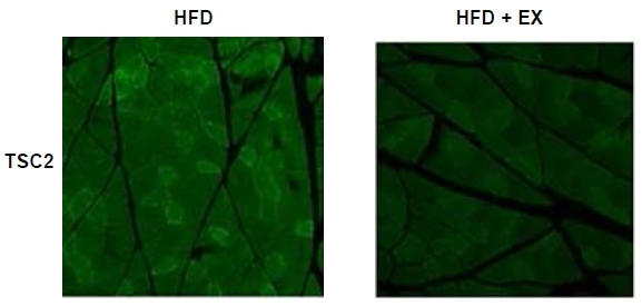
Aerobic exercise decreases TSC2 protein expression in the plantaris muscles of high-fat-fed OVX rats
To test the effect of the high-fat diet and aerobic exercise on total TSC2 protein expression, which inhibits the mammalian target of rapamycin (mTOR) and p70S6K, we performed immunohistochemical staining. As expected, TSC2 (green) was strongly detected in the HFD group. Conversely, aerobic exercise largely eliminated the high-fat-induced increase in TSC2 expression (Fig. 1).
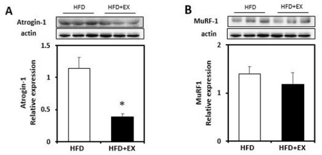
Effect of aerobic exercise on ubiquitin ligases (atrogin-1 and MuRF1) protein and mRNA expression in the skeletal muscles of high-fat-fed OVX rats
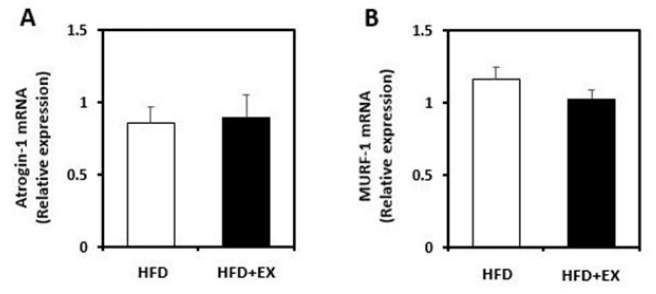
To investigate the effects of aerobic exercise on the ubiquitin ligases, we performed western blot analysis. The muscle content of atrogin-1 was significantly decreased in the HFD+EX group (p< 0.05; Fig. 2A). Although the difference was not statistically significant, MuRF1 also tended to decrease in the exercise group when compared with the HFD group. As shown in Fig. 4A, real-time qPCR analysis revealed that atrogin-1 gene expression was not significantly decreased in the exercise group when compared with the non-exercise group. The MuRF1 mRNA level was also not significantly decreased in the exercise group. (Fig. 4B).
Figure┬Ā2.
Immunoblot analyses of protein abundance of (A) Atrogin-1, and (B) MuRF-1 in plantaris muscles from ovariectomized rats fed a high-fat diet (HFD), or a high-fat diet with aerobic exercise for 8 weeks (HFD+EX). Target protein values are shown normalized to the actin expression level for each sample. (n=3/group; mean ┬▒ SE; *p< 0.05).
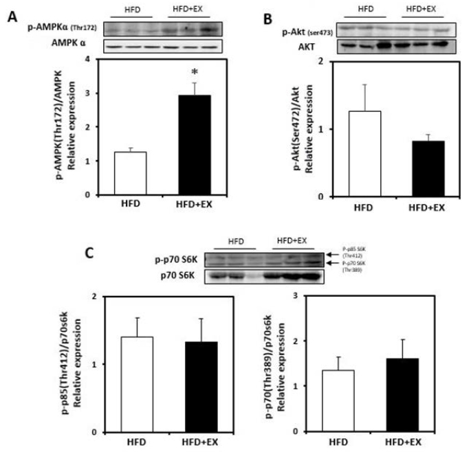
Figure┬Ā3.
Immunoblot analyses of protein abundance of (A) phosphorylation of Thr172 on AMPK, (B) phosphorylation of Ser473 on Akt, and (C) phosphorylation of Thr389 on p70S6K in plantaris muscles from ovariectomized rats fed a high-fat diet (HFD), or a high-fat diet with aerobic exercise for 8 weeks (HFD+EX). Target protein values are shown normalized to each protein expression level for each sample. (n=3/group; mean ┬▒ SE; *p< 0.05).
Figure┬Ā4.
Real-time qPCR was performed to assess mRNA level of plantaris muscles on ovariectomized rats fed a high-fat diet (HFD), or a high-fat diet with aerobic exercise for 8 weeks (HFD+EX). (A) Atrogin-1, and (B) MuRF-1 mRNA levels. All results are normalized to GAPDH mRNA level for each sample. (n=9/group; mean ┬▒ SE).
Effect of aerobic exercise on AMPK, AKT, and p70S6K phosphorylation in the high-fat-fed OVX rat plantaris muscle
The phosphorylation of AMPK (Thr172) was significantly increased in response to aerobic exercise (p<0.05; Fig. 3A). However, the p-AKT (Ser473) activity was not altered in the exercise group (Fig. 3B). To demonstrate whether AMPK mediates the inhibition of the mTOR signaling pathway, we measured the level of p70S6K protein activity. As shown in Fig. 3C, p70S6K protein phosphorylation was not significantly elevated in the exercise group.
DISCUSSION
The efficiency of maintaining skeletal muscle mass declines with age. The age-related loss of estrogen production in menopause promotes increased lipogenesis and decreased skeletal muscle mass. It is well recognized that resistance exercise training promotes skeletal muscle protein synthesis through the phosphatidylinositol 3-kinase (PI3-k)-Akt-mTOR cascade7, 20, 21, and decreasing the muscle-specific ubiquitin ligases. However, aerobic exercise has a negligible effect on skeletal muscle mass regulation. In fact, aerobic exercise is commonly recommended for improvements of cardio-metabolic health, respiratory fitness and cardiovascular function through the activation of AMPKPGC-1╬▒ signaling pathways20. muscle protein degradation via suppression of TSC2, independent of AMPK activity, but p70S6K and mTOR downstream target activation are not regulated through low-intensity aerobic exercise.
It is well known that fat accumulation and skeletal muscle weakness ensue when estrogen levels decline with age in women. Both type I and type II muscle fibers are smaller in women than men and type II fibers are more affected with aging than type I fibers22, 23. Therefore, the current paradigm is that resistance exercise training is a necessity for type II fiber mass in post-menopausal women. Aerobic exercise is a conventional exercise prescription to induce skeletal muscle aerobic capacity and metabolic improvement; however, the purpose of our study was to examine the effect of low-intensity aerobic exercise training on type II-rich skeletal muscle mass in the high-fat-fed ovariectomized rat. Because low-intensity aerobic exercise is an easier and safer method than resistance exercise, it is better suited for post-menopausal elderly women. Here, we report that decreased protein expression of skeletal muscle atrophy markers were observed in plantaris muscles of high-fat-fed OVX rat following 8 weeks of low-intensity aerobic exercise training. Therefore, our study suggests that aerobic exercise can lead to reduced rates of protein degradation in the type II skeletal muscle fiber. However, skeletal muscle hypertrophy pathways were not activated through low-intensity aerobic exercise in the plantaris muscle.
CONCLUSION
In conclusion, our results suggest that 8 weeks of low-intensity aerobic exercise training induced decremental changes in atrogin-1 and TSC2 protein expression in the plantaris muscles of high-fat-fed ovariectomized rats, but activation of the Akt and mTOR downstream pathways were not observed. Based on these results, we propose that low-aerobic exercise should be considered as an important component of recommendations in combatting skeletal muscle atrophy that occurs with menopause.


