


INTRODUCTION
In the field of music therapy, lyrical music with a stable beat and volume is generally used for psychotherapy because it induces mentally positive effects [1,2]. In contrast, High-Intensive Sound (HIS) including music could be recognized as a noise by some individuals. Music in general is played around 85 dB, rock bands with loud sounds play around 110 dB, and a jet engine's noise is around 150 dB. HIS of 120 dB or higher can cause a painful listening experience, and hearing impairment occurs if one is exposed continuously to a noise of 80 dB or higher. It has been reported that the sound of 96-110 dB can cause various negative effects on the body depending on the duration of the exposure [3]. As such, sound can affect the body either positively or negatively based on the volume.
High-decibel music (HDM) can be interpreted as noisy or joyful based on one's tolerance. The hedonic qualities of HDM are difficult to measure as they are subjective. HDM played during sports or exercise induces excitement, and it is occasionally used to maintain performance. However, despite countless research reports on brain activation by exercise, there have been hardly any studies on cognitive functions and brain activity stimulated by exercise combined with HDM.
Studies on the relationship between exercise and the brain have been actively pursued globally since 2000. Several research groups in Korea have demonstrated neurogenesis in the hippocampus and improved short-term memory in rodents in response to exercise [4-7]. Few studies in humans have assayed improvements in cognitive functions due to exercise. These studies have used the Stroop test [8] and the cerebral blood flow velocity [9-11]; however, none have looked at the activation of the brain and changes in oxygenated hemoglobin (Oxy-Hb). Various molecular biology and behavior studies have been conducted that found learning and memory increases in response to augmented neurogenesis in the hippocampus and a decreased time to find the platform in the Morris water maze task [12]. In humans, long-term medium intensity exercise resulted in memory improvements combined with decreased selection-reaction time, increased brain size, prefrontal cortex activation, and the improvements in cognitive function [13-15]. While studying the relationship between exercise and cerebral blood oxygenation using near infrared spectroscopy (NIRS), it has been observed that acute exercise increases Oxy-Hb of the prefrontal cortex, and that young people and the old people have different areas of brain activation during exercise [16,17]. Many aspects regarding the relation between brain activation and an age group, exercise intensity, exercise type, or surrounding environment remain to be explored.
NIRS, a method for assessing brain activation, is a non-invasive measurement that can monitor metabolic function by assessing blood flow and oxygen saturation using light wavelengths between 700-3,000 nm. NIRS can measure information about activation in a particular brain area by measuring the concentration changes in Oxy-Hb and deoxygenated hemoglobin (Deoxy-Hb) within the body (the brain or muscle). The biggest strength of NIRS, compared to other brain imaging methods, is that real-time assessment of exercise is possible as long as there are no drastic movements of the head such as in cycling. When performing a cognitive activity or exercise, neurons in the corresponding brain areas are activated, and they require oxygen supply for energy consumption. Blood flow increases to activated neurons to meet the demand. On such occasions, Oxy-Hb within the blood releases oxygen to neurons and is converted into Deoxy-Hb. To avoid a state of oxygen depletion due to oxygen consumption, an increase in Oxy-Hb concentration and decrease in Deoxy-Hb concentration occurs. The Stroop test is commonly used to measure prefrontal cortex activation. NIRS has been used to confirm that the Stroop test measures prefrontal cortex activation by detecting significant augmentation in Oxy-Hb in bilateral dorsolateral prefrontal cortex (DLPFC) during the task [16]. The prefrontal cortex is involved in thought, behavioral control, speech, decision making, emotional control, memory control, concentration, distraction, and motivation. Humans recognize the meaning of a letter subconsciously, and thus it is difficult to determine the letter and its color at the same time. This is called cognitive interference (Stroop interference), and it reflects the selection function of the prefrontal cortex [18-20]. Studies measuring cerebral blood flow during the Stroop task have demonstrated that patients with vascular dementia display poor performance on the Stroop task compared to healthy people because of reduced blood flow to the DLPFC area [21-23]. As the Stroop task requires prefrontal cortex activation, this simple test can infer the activation of brain function in the prefrontal cortex.
Several studies have reported that medium-intensity exercise increases the activation of the frontal lobe. However, people who exercise for health, including athletes, often perform high-intensity exercise combined with HDM to induce “excitement and fun.” Nevertheless, there have been few studies on the effects of listening to HDM during high-intensity exercise on cerebral activation.
Therefore, we studied blood flow changes to the prefrontal cortex using the Stroop test and NIRS in order to understand the effects of a combination of HDM and high-intensity exercise on the prefrontal cortex. This study seeks to explore the effects of being exposed to HDM during exercise on cognitive function.
METHODS
Subjects
The subjects of this study were 28 female university students. The study subjects did not have any particular disease and they were drug-free, healthy individuals, and such selection was done so that they can handle exercise stress of cycle ergometer without difficulties. They were categorized into 4 groups, Control Group (CG), Music Group (MG), Exercise Group (Ex), Music and Exercise Group (MnEx), and the study was conducted with a crossover design so that all the subjects were able to participate in all the 4 groups. All the subjects had a total of 5 tests including a maximal exercise stress test. The characteristics of subjects are listed in Table 1.
Maximal Oxygen Uptake Test
Maximal oxygen uptake was assessed with a gas analyzer in order to set equal exercise intensity relative to each subject's capacity (MediGraphic, VO2000, The U.S.). Regarding an assessment method protocol, warm up of 1 minute was done using cycle ergometer (Ergomedic 839E, Monark co., Sweden) and then started at 30W and increased 25W per every 2 minutes (Fig. 1). VO2 max and heart rate values from this test were used to set exercise stress.
Stroop and NIRS test (Cognitive function)
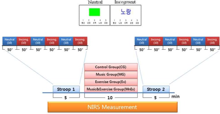
We attached the sensor of NIRS equipment (Hamamatsu, NIRO-200) onto the prefrontal cortex area of the test subject, and had the subject sit on cycle ergometer and let her rest until the heart rate was stabilized on a wireless heart rate monitor. After the rest, 5 minutes of Stroop test was conducted with computer program. For a Stroop test method, 5 seconds was set for one question, and the time required to answered the question was assessed per millisecond. For a question method, it was processed alternatively with 1~10 being neutral, 11~20 being incongruent, 21~30 being neutral, 31~40 being incongruent, 41~50 being neutral and 51~60 being incongruent. 5 examples were given per question and the reaction time of pushing a computer key was assessed. Questions were comprised differently based on the test order of researchers per each session (Fig. 2).
Experimental Procedure
All the stages of experiment were conducted as follows. 1) Analyzing height, weight and body fat (Inbody 7.0, Biospace) and getting consent for the experiment. 2) Having the subject sit on cycle ergometer, attaching NIRS sensor onto the prefrontal cortex area (left forehead) and putting on a wireless heart rate monitor. 3) After taking a rest, conducting 5 minutes of pre-Stroop test using computer program. 4) Conducting intervention for 10 minutes (CG: rest, MG: listening to the music of 100dB or higher, Ex: cycle exercise with 70-75% of VO2 max, MnEx: MG+Ex) → post-Stroop test. 5) During the entire process, Oxy-Hb, Deoxy-Hb, Tissue Oxygen Index (TOI: showing oxygen saturation) and normalized Tissue Hemoglobin Index (nTHI: indicator of hemoglobin to tissue) were assessed in real time with NIRS sensor, attached onto the prefrontal cortex.
Statistical analysis
All the result of this study was listed in Mean ± SD and IBM Statistics 20 was used for all the statistical analysis of this study. One-way ANOVA was conducted to understand differences based on each treatment, and post hoc test was conducted with Fisher’s LSD (Least Significant Difference) for the variables which showed a significant difference. For independent measurement variables, paired t-test was conducted to verify them. The significance level of 0.05 was used for all test.
RESULTS
Maximal Oxygen Uptake Test
The results obtained with maximal exercise stress test, using cycle ergometer, are listed in <Table 2>. For the test subjects, it took 8 minutes and 34 seconds from maximal exercise stress test to All-Out. Average maximum heart rate was 171.8 ± 15.4 times/minute and maximal oxygen uptake was 29.2 ± 4.3 ml/kg/min.
Stroop test (Cognitive function)
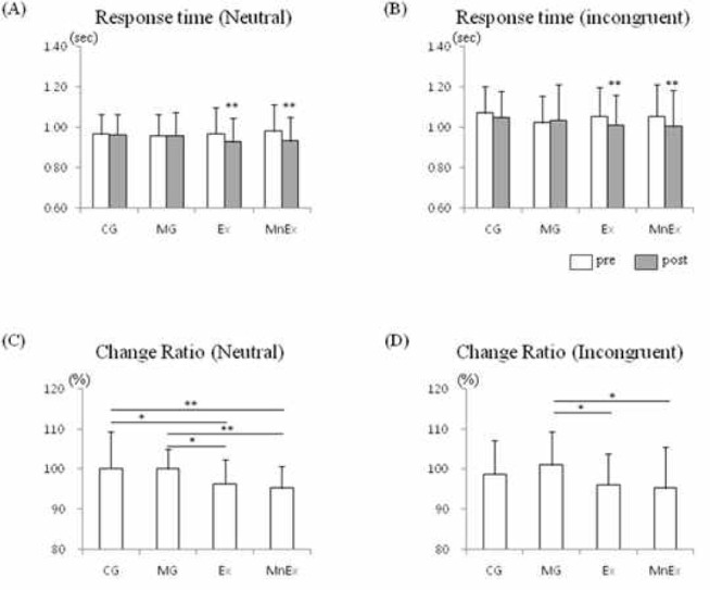
Neutral and incongruent reaction time, which were the results of pre & post-Stroop test, are listed in <Fig. 3>. From the results of both situations, there was no significant difference between before and after the treatment for CG and MG. However, for both of Ex and MnEx, faster reaction time was shown in post-Stroop compared to pre-Stroop (p < 0.01, both). In the neutral task, the change rate from pre-Stroop to post-Stroop was significantly decreased in Ex (p < 0.05) and MnEx (p < 0.01) compared to CG, and also significantly decreased in Ex (p < 0.05) and MnEx (p < 0.01) compared to MG. From the incongruent task, a significant decrease occurred in Ex (p < 0.05) and MnEx (p < 0.05) compared to MG. And there was no significant difference when compared to CG.
The prefrontal cortex of the brain activation
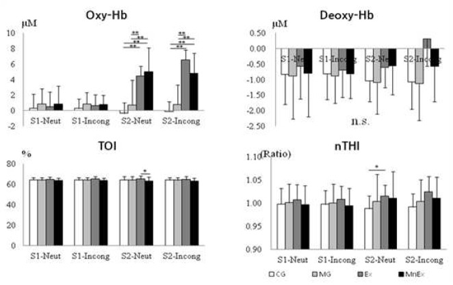
The change per each group on Oxy-Hb, Deoxy-Hb, TOI and nTHI in the prefrontal cortex during the stages of this experiment are listed in <Fig. 4> in graphs. Oxy-Hb and Deoxy-Hb had drastic increase after the treatment in Ex and MnEx. Both of groups did not have big decrease of Oxy-Hb during Stroop test after the exercise. Compared to other groups, CG and MG did not have drastic change. TOI changes in all the groups were not big but in MnEx, the score was somewhat low. Regarding the change of nTHI, increase was shown in Ex and MnEx thanks to the effect of exercise. CG and MG, which did not have exercise, had decrease after the treatment.
<Fig. 5> is the change of Oxy-Hb and Deoxy-Hb per each stage of pre-Stroop test (S1-Neutral, S1- Incongruent) → treat → post-Stroop test. There was no difference among the groups when conducting neutral and incongruent tasks of Stroop 1. Drastic increase of Oxy-Hb and minor increase of Deoxy-Hb were shown in both of Ex (p < 0.01) and MnEx (p < 0.01) in the treat stage. Oxy-Hb, which were increased in 2 groups, had somewhat maintained its increase during Stroop 2 (post) as well. Oxy-Hb in Ex and MnEx were significantly increased in post-Stroop test (S2-Neutral, S2- incongruent), used in this experiment, compared to CG and MG (p < 0.01, both). But there was no significant change in Deoxy-Hb.
<Fig. 6> is the result of blood flow change of the prefrontal cortex during Stroop test. Stroop tests, which were conducted before (S1) and after (S2) the treatment, were categorized into neutral and incongruent and then activation change of the prefrontal cortex was compared. From the result, there was no significant difference of Oxy-Hb between S1-Neutral and S1-Incongruent during Stroop test. But in S2-Neutral and S2-Incongruent, conducted after the treatment, had significant differences among CG-Ex (p < 0.01), CG-MnEx (p < 0.01), MG-Ex (p < 0.01) and MG-MnEx (p < 0.01). There was no statistically significant change of Deoxy-Hb in all the groups. And there was a significant decrease of TOI between Ex and MnEx (p < 0.05) during the S2-Neutral task, which was conducted after the treatment. Also there was a significant increase of nTHI between CG and Ex (p < 0.05) during the S2-Neutral task, which was conducted after the treatment.
DISCUSSION
This study was conducted to understand the effect of HDM, during performing high-intensity exercise, on the activation of the prefrontal cortex. The sound pressure of the music in decibels was set at a level where the test subjects could endure it without hearing damage. It has been shown in previous studies that exercise is an important factor for brain activation. In animal experiments, it has been reported that neurogenesis is stimulated in the hippocampus, memory function is increased, and brain-derived neurotrophic factor expression is increased in response to exercise [12,24,25]. Compared to animal studies, studies on brain activation in humans are limited due to technical complications in analysis and assessment. In order to analyze the effect of exercise on human brains, changes in brain waves or blood flow is measured before and after exercise. These studies have demonstrated that long-term aerobic exercise of medium intensity results in activation of the prefrontal cortex, increase in cognitive function, better task performance, faster decision making, increase in brain proteins related to memory and cognitive function, and an increase in white matter volume [13,26-29]. Indeed, exercise is considered effective for the activation of brain areas involved in cognitive functions including learning and memory. Most studies on exercise and cognitive function have dealt with long-term medium intensity exercise. Recently, researchers reported brain activation due to acute exercise based on neurovascular coupling theory. Two previous studies [16,30] demonstrated through NIRS testing of changes to blood flow during exercise that one-off exercise also improves cognitive function that correlates with the activation of the frontal lobe.
Previous studies investigating the positive effects of exercise on cognition demonstrated that medium-intensity exercise, around 50% of maximal oxygen uptake, has the highest effects on mental function [14]. Many aspects of the effects of exercise on cognitive function based on the environment are yet to be investigated. In this study, stationary cycles, which offer easy assessment during exercise, were used to analyze the effect of HDM combined with high-intensity exercise on cognitive reaction time and blood flow to the prefrontal cortex in real time.
We conducted preliminary studies on the effect of HDM combined with high intensity exercise on the Stroop score. We found that the Stroop score was lower in the HDM plus high-intensity exercise group than in the HDM only or high-intensity exercise only group [31]. In this study, we attempted to prove in brain blood flow flow-up on these findings; however, we found no significant difference in reaction time on the Stroop task (Fig. 3). When Stroop scores were compared before and after the treatment, performing exercise was found to results in quicker reaction time compared to average response time. The number of wrong answers after the treatment was not statistically significant compared to the number before the treatment. This finding is identical with a previous study where wrong answer rate remained the same while reaction time decreased [29]. Whether HDM was included or not, exercise brought significant improvement in reaction time on the Stroop task for both neutral and incongruent tasks. However, only the Ex and MnEx compare to MG showed statistically significant changes in the incongruent task when comparing the extent of changes before and after the task (Fig. 3D). These results suggest that HDM can affect cognitive function when performing a complex task.
Blood flow changes to the prefrontal cortex was assessed in this study in real time for the entire length of the test, including exercise. We found that exercise activates the prefrontal cortex during and even after a session (Fig. 5 & Fig 6). We found no difference among groups during the pre-Stroop task since all subjects participated in all tests (Fig. 5). We found a significant increase in Oxy-Hb and Deoxy-Hb in the Ex and MnEx groups after the treatment. However, there was no difference between the two groups supporting the findings of the Stroop task. Oxy-Hb concentration during the Post-Stroop task was identical with that after-treatment, and there was no significant difference in Deoxy-Hb. The increase in Deoxy-Hb concentration only right after the treatment could be due to intense cycling exercise (70-75% of VO2 max). It is highly plausible that the decrease in reaction time was due to an increase in alertness from the neural activity-promoting effects of exercise [32]. In addition, it was previously reported that changes in catecholamine levels due to exercise cause changes in alertness [33].
We next measured changes in blood flow during intense pre- and post-Stroop tasks (Fig 6) Oxy-Hb increased in Ex, MnEx compare to CG and Ex, MnEx compare to MG only during the post-Stroop task. We found no difference in Deoxy-Hb among groups during all the tasks. These findings are consistent with the Stroop reaction time results. However, TOI, measured by blood-to-tissue ratio of Oxy-Hb, showed a significant decrease in MnEx than in Ex during the post-Stroop (neutral) task. These findings suggest that exercise with HDM can cause a negative effect on prefrontal cortex activation. In addition, we found a significant increase in nTHI only in the CG-Ex group during the post-Stroop (neutral) task. These result show that HDM with or without exercise did not increase nTHI; exercise only had a positive effect on the prefrontal cortex activation.
CONCLUSION
In this study, we analyzed the effects of high intensity exercise combined with HDM on cognitive function and blood flow to the prefrontal cortex. Exercise decreases the reaction time during the Stroop task, and at the same time increases Oxy-Hb in the prefrontal cortex. This study provides evidence that exercise improves cognitive function through prefrontal cortex activation. However, 10 min of HDM (100 dB) during exercise did not affect cognitive function. Interestingly, we found that the TOI of the prefrontal cortex decreased in MnEx than in Ex, and nTHI significantly increased only in Ex. This suggests that HDM possibly has negative effects on prefrontal cortex activation.